CELL COMMUNICATION

 Brief table of contents 

    What is Signaling?

    Have you ever become separated from a friend while in a crowd? 
    You know the challenge of Finding someone when surrounded by hundreds of other people. If you and your friend have Mobile phones, your chances of finding each other are very good. A cell phone’s ability to send and receive messages makes it an ideal communication device.

    Cell Signaling

    In biology, cell signaling ( cell signaling in British English ), or cell-cell communication, controls the basic actions of cells and coordinates actions with multiple cells. Signals an object that encodes or conveys information. Biological processes are complex molecular interactions that involve many signals. The ability of cells to perceive and properly respond to their microenvironment is the basis of development, tissue repair, and immunity. like normal tissue homeostasis. Errors in signaling interactions and cellular information processing can cause diseases such as cancer, autoimmunity, and diabetes. By understanding cell signaling, doctors can treat diseases more effectively, and in theory, researchers could create artificial tissues.

    Systems biology studies the basic structure of cell signaling networks and how changes in these networks can affect the transmission and flow of information ( signal transduction ). Such networks are complex systems in their organization and may exhibit a number of emergent properties, including bistability and hypersensitivity. The analysis of cell signaling networks requires a combination of experimental and theoretical approaches, including the development and analysis of simulations and simulations. Far allostery is often an important component of cell signaling events.

    Signaling agents can be physical agents such as mechanical pressure, voltage, temperature, light, etc., or chemical agents such as peptides, steroids, terpenoids, etc. Simply put, each molecule has some or some signaling consequences. it could be food material or pathogen-associated structures, or it could be oxygen levels or carbon dioxide, or it could be specially biosynthesized signaling molecules such as hormones and pheromones (ectohormones). Signal molecules vary greatly in their physicochemical properties such as solubility (hydrophobic or hydrophilic). Some signaling molecules are gaseous, such as nitric oxide.

    All cells receive signals from their environment and respond to them. This is achieved by multiple signaling molecules that are secreted or expressed on the surface of one cell and bind to a receptor expressed by other cells, thereby integrating and coordinating the function of the many individual cells that make up organisms. Each cell is programmed to respond to certain extracellular signaling molecules. Extracellular signaling typically involves the following steps:

    1. Synthesis and release of the signaling molecule by the signaling cell ;
    2. Signal transport to the target cell ;
    3. Binding of a signal to a specific receptor, leading to its activation ;
    4. Initiation of signal transduction pathways.
    Synthesis involves various biosynthetic pathways and occurs at a specific time and place. Sometimes signaling molecules are released from the cell, and sometimes they are not released at all, such as cellular localization signals and DNA damage signals. Such intracellular signaling networks operate within the cell. Sometimes, signaling molecules are released via various pathways such as membrane diffusion, exocytosis, cell rupture, etc. In some cases, signaling molecules remain attached to the cell surface, a mode that aids in juxtacrine signaling (discussed below). Sometimes signaling molecules require activation, such as through proteolytic cleavage, covalent modification, etc.

    The signal pathway can be intracellular or intercellular. Intercellular signaling is also referred to as intercellular communication. It can be short or long distance. Based on the nature of this signal molecule pathway from source to target cell; signaling pathways are categorized into autocrine, juxtacrine, intracrine, paracrine, and endocrine.

    Receptors play a key role in cell signaling. Receptors help recognize the signaling molecule (ligand). However, some receptor molecules interact with a physical agent such as voltage, light, etc. Receptor molecules are usually proteins, but in some cases, they may be RNA. Receptors can be located on the cell surface or within the cell, for example, in the cytosol, organelles, and nuclei (especially transcription factors). Normally, cell surface receptors bind membrane-impermeable signaling molecules, but sometimes they also interact with membrane-permeable signaling molecules. The key step in signal transduction is the removal and degradation of the signaling molecule. Sometimes the receptor is also degraded. neurotransmitter reuptake is a mechanism for removing signaling molecules that is commonly seen in the nervous system and is the target of some classes of prescription psychiatric drugs.

    Binding to a ligand causes a conformational change in the receptor, which leads to further signaling. Due to a conformational change, a receptor can exhibit either an enzymatic activity (called an enzyme receptor) or an ion channel opening or closing activity (called a ligand-gated channel receptor). Sometimes the receptors themselves do not contain any enzyme or channel domain but are associated with an enzyme or a carrier. Some receptors (nuclear-cytoplasmic superfamily) have a different mechanism: once they bind to a ligand, they change their DNA binding properties and cellular localization properties.

    The result of the enzymatic activity of the receptor usually leads to the recruitment of another molecular change, which again causes another molecular change, thus causing a "cascade" of signal transduction. These intermediates often form a secondary messaging system. Within the signal transduction cascade, there may be enzymes and transporters that work similarly to receptors. These enzymatic activities include covalent modifications such as proteolytic cleavage, phosphorylation/dephosphorylation, methylation/demethylation, ubiquitinylation/deubiquitinylation, etc. They are part of regulatory strategies. Reactions take place at very specific sites on the substrate. An important phenomenon that occurs in the intracellular part of signaling is signal amplification. through which, even if a small number of receptors are activated, a very large number of molecules in the downstream are subsequently activated. Inhibition and activation (through feed-forward or feedback) and crosstalk are also important phenomena in signal chains. Sometimes the feedback causes biological fluctuations. With prolonged exposure or overexposure to a signal, a mechanism called adaptation or desensitization occurs, whereby the additional signal is ignored. Reactivation of resensitization may occur after the extra stimulus has been removed for an extended period.

    Between separate organisms of the same species

    Cell signaling has been most extensively studied in the context of human diseases and signaling between cells of the same organism. However, intercellular signaling can also occur between cells of two different individuals of the same species. In many mammals, early embryonic cells exchange signals with uterine cells. In the human gastrointestinal tract, bacteria communicate with each other and with the human epithelium and cells of the immune system. For the yeast Saccharomyces cerevisiae, during mating, some cells send a peptide signal( pheromone mating factor ) into their environment. The mating factor peptide can bind to a receptor on the cell surface of other yeast cells and induce them to prepare for mating.

    Example:

    Signaling in bacteria enables bacteria to monitor extracellular conditions, ensure that there are sufficient amounts of nutrients, and ensure that hazardous situations are avoided. There are circumstances, however, when bacteria communicate with each other.

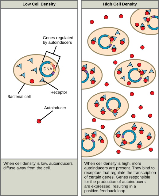
    The first evidence of bacterial communication was observed in a bacterium that has a symbiotic relationship with Hawaiian bobtail squid. When the population density of the bacteria reaches a certain level, specific gene expression is initiated, and the bacteria produce bioluminescent proteins that emit light. Because the number of cells present in the environment (cell density) is the determining factor for signaling, bacterial signaling was named quorum sensing. In politics and business, a quorum is the minimum number of members required to be present to vote on an issue.

    Quorum sensing uses autoinducers as signaling molecules. Autoinducers are signaling molecules secreted by bacteria to communicate with other bacteria of the same kind. The secreted autoinducers can be small, hydrophobic molecules such as acyl-homoserine lactone, (AHL) or larger peptide-based molecules; each type of molecule has a different mode of action. When AHL enters target bacteria, it binds to transcription factors, which then switch gene expression on or off (see Figure 1). The peptide autoinducers stimulate more complicated signaling pathways that include bacterial kinases. The changes in bacteria following exposure to autoinducers can be quite extensive. The pathogenic bacterium Pseudomonas aeruginosa has 616 different genes that respond to autoinducers.

    Signaling in bacteria

    Some species of bacteria that use quorum sensing form biofilms, complex colonies of bacteria (often containing several species) that exchange chemical signals to coordinate the release of toxins that will attack the host. Bacterial biofilms (Figure 2) can sometimes be found on medical equipment; when biofilms invade implants such as hip or knee replacements or heart pacemakers, they can cause life-threatening infections.

    Cell-cell communication enables Staphylococcus aureus bacteria (Figure 2a) to work together to form a biofilm inside a hospital patient’s catheter, seen here via scanning electron microscopy. S. aureus is the main cause of hospital-acquired infections. Hawaiian bobtail squid (Figure 2b) have a symbiotic relationship with the bioluminescent bacteria Vibrio fischeri. The luminescence makes it difficult to see the squid from below because it effectively eliminates its shadow. In return for camouflage, the squid provides food for the bacteria. Free-living V. fischeri do not produce luciferase, the enzyme responsible for luminescence, but V. fischeri living in a symbiotic relationship with the squid do. Quorum sensing determines whether the bacteria should produce the luciferase enzyme.


    (a) Staphylococcus aureus bacteria. (b) Hawaiian bobtail squid. (credit a: modifications of work by CDC/Janice Carr; credit b: modifications of work by Cliff1066/Flickr)
    Research on the details of quorum sensing has led to advances in growing bacteria for industrial purposes. Recent discoveries suggest that it may be possible to exploit bacterial signaling pathways to control bacterial growth; this process could replace or supplement antibiotics that are no longer effective in certain situations.

    Classification

    Cell signaling can be defined as either mechanical or biochemical depending on the type of signal. Mechanical signals are the forces acting on the cell and the forces generated by the cell. These forces can be perceived by cells and react to them. Biochemical signals are biochemical molecules such as proteins, lipids, ions, and gases. These signals can be categorized according to the distance between the signaling and responding cells. Signaling within, between and between cells is classified into the following classifications:

    • Intracrine signals are produced by the target cell, which remains within the target cell. See More
    • Autocrine signals are produced by the target cell, secreted, and act on the target cell itself.  through receptors. Sometimes, autocrine cells can target nearby cells if they are the same type as the emitting cell. An example of this is immune cell signals. See More
    • Juxtacrine is directed to neighboring (adjacent) cells. These signals are transmitted along cell membranes through protein or lipid components embedded in the membrane and are able to act either on the emitting cell or directly adjacent cells. See More
    • Paracrine signals to target cells in the vicinity of the emitting cell. Neurotransmitters are an example. See More
    • Endocrine signals target distant cells. Endocrine cells produce hormones that travel through the bloodstream to reach all parts of the body. See More

    Some cell-to-cell communication requires a direct cell-to-cell contact . Some cells can form gap junctions , which connect their cytoplasm to that of neighboring cells. In cardiac muscle , gap junctions between neighboring cells propagate an action potential from the pacemaker region to propagate and coordinate the contraction of the heart.

    The notch signaling mechanism is an example of juxtacrine signaling (also known as contact-dependent signaling), in which two neighboring cells must make physical contact in order to communicate. . This direct contact requirement allows very precise control of cell differentiation.during embryonic development. In the worm Caenorhabditis elegans, the two cells of the developing gonad have an equal chance of finally differentiating or becoming a uterine progenitor cell that continues to divide. The choice of which cell continues to divide is controlled by the competition of cell surface signals. One cell will produce more cell surface protein that activates the Notch receptoron the next cell. This activates a feedback loop or system that reduces Notch expression in the cell that will differentiate and that increases Notch on the cell surface that continues as a stem cell.
    Notch-mediated juxtacrine signal between neighboring cells


    Many cellular signals are carried by molecules that are released by one cell and move to make contact with another cell. Endocrine signals are called hormones. Hormones are produced by endocrine cells, and they travel through the bloodstream to reach all parts of the body. The specificity of signaling can be controlled if only some cells can respond to a certain hormone. Paracrine signals, such as retinoic acid , only target cells in the vicinity of the emitting cell. neurotransmitters present another example of a paracrine signal. Some signaling molecules can function as both a hormone and a neurotransmitter. For example, epinephrine and norepinephrine can act as hormones when released from the adrenal glands and transported to the heart through the bloodstream. Norepinephrine can also be produced by neurons in order to function as a neurotransmitter in the brain. Estrogen can be secreted by the ovary and act as a hormone or act locally via paracrine or autocrine signaling. Reactive oxygen and nitric oxide speciesmay also act as cellular messengers. This process is called redox signaling.

    In Multicellular Organisms

    In a multicellular organism, signaling between cells occurs either through release into the extracellular space, divided into paracrine. signaling (short distance) and endocrine signaling (long distance) or by direct contact, known as juxtacrine signaling. autocrine signaling is a special case of paracrine signaling where the secreting cell has the ability to respond to the secreted signaling molecule. Synaptic signaling is a special case of paracrine signaling (for chemical synapses ) or juxtacrine signaling (for electrical synapses ) between neurons and target cells. Signaling molecules interact with the target cell as a ligandto cell surface receptors and/or by entering the cell through its membrane or endocytosis for intracrine signaling. This usually results in the activation of second messengers, leading to various physiological effects.

    A particular molecule is usually used in various signaling modes, and therefore classification by signaling modes is not possible. At least three important classes of signaling molecules are widely known, although not exhaustive and with imprecise boundaries, as such membership is not exclusive and depends on the context:

    Hormones are the primary signaling molecules of the endocrine system , although they often regulate each other's secretion through local signaling (e.g. islet cells of Langerhans ), and most are also expressed in tissues for local purposes (e.g. angiotensin ) or otherwise structurally related molecules (e.g. PTHrP ).
    neurotransmitters are the signaling molecules of the nervous system, including also neuropeptides and neuromodulators. Neurotransmitters such as catecholamines are also secreted by the endocrine system into the systemic circulation.
    Cytokines are signaling molecules of the immune system, with a primary paracrine or juxtacrine role, although during significant immune responses they may have a strong presence in the circulation with a systemic effect (altering iron metabolism or body temperature ). Growth factors can be considered as cytokines or another class.
    Signal molecules can belong to several chemical classes: lipids, phospholipids , amino acids , monoamines, proteins, glycoproteins, or gases. Signaling molecules that bind surface receptors are usually large and hydrophilic (e.g., TRH, vasopressin, acetylcholine ), while those that enter the cell are usually small and hydrophobic (e.g., glucocorticoids , thyroid hormones, cholecalciferol ,retinoic acid ), but important exceptions to both are numerous, and the same molecule can act both through a surface receptor and in an intracrine manner, producing different effects. In intracrine signaling, once inside the cell, the signaling molecule can bind to intracellular receptors , other elements, or stimulate enzyme activity (such as gases). The intracrine action of peptide hormones remains a matter of controversy.

    Hydrogen sulfide is produced in small amounts by some cells in the human body and has a number of biological signaling functions. Currently, only two other such gases are known to act as signaling molecules in the human body: nitric oxide and carbon monoxide .

    In plants

    Signaling in plants occurs through plant hormones, phytochromes, cryptochromes, etc.

    Important families of plant hormones are auxin, cytokinin, gibberellin, ethylin, jasmonic acid, salicylic acid, strigolactones, polyamines, nitric oxide, peptide hormones, etc. RNA translocation has also been reported.

    Cell To Cell Signaling in plants

    Cells typically communicate using chemical signals. These chemical signals, which are proteins or other molecules produced by a sending cell, are often secreted from the cell and released into the extracellular space. There, they can float – like messages in a bottle – over to neighboring cells.


    Cell To Cell Signaling in plants

    Not all cells can “hear” a particular chemical message. In order to detect a signal (that is, to be a target cell), a neighbor cell must have the right receptor for that signal. When a signaling molecule binds to its receptor, it alters the shape or activity of the receptor, triggering a change inside of the cell. Signaling molecules are often called ligands, a general term for molecules that bind specifically to other molecules (such as receptors).
    The message carried by a ligand is often relayed through a chain of chemical messengers inside the cell. Ultimately, it leads to a change in the cell, such as alteration in the activity of a gene or even the induction of a whole process, such as cell division. Thus, the original intercellular (between-cells) signal is converted into an intracellular (within-cell) signal that triggers a response.


    Cell To Cell Signaling in plants 2
    Cell-cell signaling involves the transmission of a signal from a sending cell to a receiving cell. However, not all sending and receiving cells are next-door neighbors, nor do all cell pairs exchange signals in the same way.
    There are four basic categories of chemical signaling found in multicellular organisms: paracrine signaling, autocrine signaling, endocrine signaling, and signaling by direct contact. The main difference between the different categories of signaling is the distance that the signal travels through the organism to reach the target cell.

    Paracrine signaling

    Often, cells that are near one another communicate through the release of chemical messengers (ligands that can diffuse through the space between the cells). This type of signaling, in which cells communicate over relatively short distances, is known as paracrine signaling.
    Paracrine signaling allows cells to locally coordinate activities with their neighbors. Although they're used in many different tissues and contexts, paracrine signals are especially important during development, when they allow one group of cells to tell a neighboring group of cells what cellular identity to take on.

    Synaptic signaling

    One unique example of paracrine signaling is synaptic signaling, in which nerve cells transmit signals. This process is named for the synapse, the junction between two nerve cells where signal transmission occurs.
    When the sending neuron fires, an electrical impulse moves rapidly through the cell, traveling down a long, fiber-like extension called an axon. When the impulse reaches the synapse, it triggers the release of ligands called neurotransmitters, which quickly cross the small gap between the nerve cells. When the neurotransmitters arrive at the receiving cell, they bind to receptors and cause a chemical change inside of the cell (often, opening ion channels and changing the electrical potential across the membrane).
    Synaptic signaling


    Synaptic signaling

    One unique example of paracrine signaling is synaptic signaling, in which nerve cells transmit signals. This process is named for the synapse, the junction between two nerve cells where signal transmission occurs.
    When the sending neuron fires, an electrical impulse moves rapidly through the cell, traveling down a long, fiber-like extension called an axon. When the impulse reaches the synapse, it triggers the release of ligands called neurotransmitters, which quickly cross the small gap between the nerve cells. When the neurotransmitters arrive at the receiving cell, they bind to receptors and cause a chemical change inside of the cell (often, opening ion channels and changing the electrical potential across the membrane).

    Signaling molecules and cellular receptors

    Autocrine signaling

    In autocrine signaling, a cell signals to itself, releasing a ligand that binds to receptors on its own surface (or, depending on the type of signal, to receptors inside of the cell). This may seem like an odd thing for a cell to do, but autocrine signaling plays an important role in many processes.
    For instance, autocrine signaling is important during development, helping cells take on and reinforce their correct identities. From a medical standpoint, autocrine signaling is important in cancer and is thought to play a key role in metastasis (the spread of cancer from its original site to other parts of the body. In many cases, a signal may have both autocrine and paracrine effects, binding to the sending cell as well as other similar cells in the area.

    Endocrine signaling

    When cells need to transmit signals over long distances, they often use the circulatory system as a distribution network for the messages they send. In long-distance endocrine signaling, signals are produced by specialized cells and released into the bloodstream, which carries them to target cells in distant parts of the body. Signals that are produced in one part of the body and travel through the circulation to reach far-away targets are known as hormones.
    In humans, endocrine glands that release hormones include the thyroid, the hypothalamus, and the pituitary, as well as the gonads (testes and ovaries) and the pancreas. Each endocrine gland releases one or more types of hormones, many of which are master regulators of development and physiology.
    For example, the pituitary releases growth hormone (GH), which promotes growth, particularly of the skeleton and cartilage. Like most hormones, GH affects many different types of cells throughout the body. However, cartilage cells provide one example of how GH functions: it binds to receptors on the surface of these cells and encourages them to divide.
    Endocrine signaling

    Signaling through cell-cell contact

    Gap junctions in animals and plasmodesmata in plants are tiny channels that directly connect neighboring cells. These water-filled channels allow small signaling molecules, called intracellular mediators, to diffuse between the two cells. Small molecules and ions are able to move between cells, but large molecules like proteins and DNA cannot fit through the channels without special assistance.
    The transfer of signaling molecules transmits the current state of one cell to its neighbor. This allows a group of cells to coordinate their response to a signal that only one of them may have received. In plants, there are plasmodesmata between almost all cells, making the entire plant into one giant network.
    Signaling molecules and cellular receptors


    In another form of direct signaling, two cells may bind to one another because they carry complementary proteins on their surfaces. When the proteins bind to one another, this interaction changes the shape of one or both proteins, transmitting a signal. This kind of signaling is especially important in the immune system, where immune cells use cell-surface markers to recognize “self” cells (the body's own cells) and cells infected by pathogens.

    Adaptive immune response

    Signaling in plants occurs through plant hormone

    Plant growth and development are regulated by a structurally unrelated collection of small molecules called plant hormones. During the last 15 years, the number of known plant hormones has grown from five to at least ten. Furthermore, many of the proteins involved in plant hormone signaling pathways have been identified, including receptors for many of the major hormones. Strikingly, the ubiquitin-proteasome pathway plays a central part in most hormone-signaling pathways. In addition, recent studies confirm that hormone signaling is integrated at several levels during plant growth and development.  

    Because plants have a sessile lifestyle, they must adjust to numerous external stimuli and coordinate their growth and development accordingly. The plant hormones, a group of structurally unrelated small molecules, are central to the integration of diverse environmental cues with a plant’s genetic program. The ‘classical’ phytohormones, identified during the first half of the twentieth century, are auxin, abscisic acid, cytokinin, gibberellin, and ethylene. More recently, several additional compounds have been recognized as hormones, including brassinosteroids, jasmonate, salicylic acid, nitric oxide, and strigolactones (Table)

    (Table 1)


    Plants also use several peptide hormones to regulate various growth responses, but this class of hormones is beyond our scope here. With the application of genetic approaches, mainly in Arabidopsis thaliana, many aspects of hormone biology have been elucidated. Most hormones are involved in many different processes throughout plant growth and development. This complexity is reflected by the contributions of hormone synthesis, transport, and signaling pathways, as well as by the diversity of interactions among hormones to control growth responses. Genetic screens resulted in the identification of many of the proteins involved in hormone signaling and the analysis of these proteins has contributed significantly to our current models of hormone action. One particularly exciting outcome is the recent identification of receptors for auxin, gibberellin, jasmonate, and abscisic acid (Fig. 1). 

    Explaination

    {BRI1 is a membrane associated receptor that cycles between the plasma membrane and endosomal compartments. The extracellular leucine-rich repeat domain binds brassinosteroids and transduces the signal through an intracellular kinase domain. GTG1 and GTG2 are GPCR-type G proteins that bind abscisic acid. They have inherent GTPase activity but also interact with the only canonical Ga subunit in Arabidopsis. PYR1/RCAR1 is a soluble ABA receptor that represses PP2C phosphatases in the presence of ABA. The cytokinin receptors CRE1, AHK2, and AHK3 are plasma-membrane associated and perceive cytokinin through their extracellular domains. Cytokinin binding triggers a phosphorylation cascade that is ultimately transmitted to response regulators in the nucleus. Like the cytokinin receptors, the known ethylene receptors are two-component regulators. All five receptors are active in the endoplasmic reticulum and transmit their signal through a common downstream component called CTR1. TIR1 and COI1 are F-box proteins that are integral components of SCF-type E3 ligases and recognize the plant hormones auxin and jasmonic acid respectively. GID1 is a nuclear-localized receptor for gibberellins. Gibberellin binding to GID1 results in the enhanced degradation of DELLA proteins.}


    Though far from complete, our improved understanding of hormone perception and signaling has allowed for comparisons between hormones. From there it is clear that some hormones (cytokinins, ethylene, and brassinosteroids) use well-characterized signaling mechanisms. On the other hand, the identification and characterization of the auxin and jasmonate receptors, as well as proteins in gibberellin signaling, have highlighted a novel mechanism for hormone perception in which the ubiquitin-proteasome pathway has a central role. In addition to these advances, the comparison of hormone signaling pathways between evolutionarily tractable members of the plant kingdom has yielded some important insights into the conservation and evolution of hormone signaling pathways. These comparisons have been facilitated by large-scale genome-sequencing projects such as those of Physcomitrella patens (moss), Selaginella (fern), Arabidopsis thaliana, and Oryza sativa (rice). For example, the moss genome (an ancient plant ancestor) encodes proteins that function in auxin, abscisic acid, and cytokinin signaling, whereas the genome of green algae does not, suggesting that these pathways emerged when plants were colonizing land. In contrast, a comparison of the moss genome with more recently diverged plant genomes suggests that signaling mechanisms for gibberellin, ethylene, and the brassinosteroids probably did not evolve until after the evolutionary split of moss and vascular plants. These observations will be expanded as additional hormone signaling components are identified and more genome sequences become available. This is an exciting time in the field of plant hormone biology because our knowledge of hormone biosynthesis, metabolism, transport, perception, signaling and response has grown exponentially over the past few years. As a result, recent reviews have been written for individual hormones covering topics from metabolism and transport to signaling. Here, we review some of the advances in plant hormone signaling. We focus on newly identified hormone receptors and the broad role of regulated protein turnover in plant hormone signaling pathways. We also discuss some of the ways that hormone pathways are integrated during plant growth and development. 

    Auxin perception by a new class of receptor 

    Auxin is crucial in regulating plant growth and development from embryogenesis through maturity. As were most hormone signaling proteins identified in plants, the auxin receptors were first found through mutant screens. In this case, the screen was for Arabidopsis seedlings with an altered response to auxin or auxin-transport inhibitors. Many of the auxin-resistant mutants identified in this way are disrupted in components of the Skp1/Cullin/F-box (SCF) ubiquitin ligases (E3) or in proteins that regulate SCF activity

    The E3 ligases are the last enzymes in the ubiquitin-protein conjugation pathway and confer specificity to the pathway. In the case of SCF-type E3 ligases, the F-box protein interacts directly with the substrate and thus determines the substrate specificity of the complex30. SCFs were first implicated in auxin signaling with the identification of an F-box protein called TIR1. Recessive mutations in TIR1 confer auxin resistance, implying that the protein is required for the degradation of negative regulators of auxin response. A key event in the characterization of the auxin-signaling pathway was the discovery that SCF(TIR1) is directly linked to auxin-regulated transcription.

    The auxin transcriptional response is controlled by two large families of transcription factors; the auxin/indole-3-acetic acid (Aux/IAA) proteins and the auxin response factors (ARFs) (of which Arabidopsis has 29 and 23 members respectively). ARFs bind the promoters of auxin-responsive genes and either activate or inhibit transcription depending on the type of ARF33. The Aux/IAA proteins bind to the ARFs through shared domains in both proteins called domains III and IV and repress auxin-regulated transcription34. Importantly, the Aux/ IAA proteins are short-lived; their degradation is promoted by auxin and dependent upon TIR1. Many gain-of-function mutations in Aux/ IAA genes have been isolated and in every case the mutations affect residues within a highly conserved region called domain II3. Biochemical studies demonstrated that domain II binds TIR1 and that this binding is enhanced by auxin. Although these results suggested a mechanism for auxin-dependent de-repression of transcription, how auxin promotes the SCFTIR1– Aux/IAA interaction remained unclear. Ultimately, TIR1 itself was shown to bind biologically active auxins directly and specifically9,11. Auxin binding to TIR1 increases the stability of the TIR1–Aux/IAA complex. Structural studies of TIR1 in the presence of auxin and a peptide encompassing domain II revealed how auxin promotes Aux/ IAA degradation. A single hydrophobic pocket on the surface of the leucine-rich repeat domain of TIR1 binds both auxin and the domain II peptide. Auxin binds to residues at the base of this pocket and contributes to binding of the Aux/IAA protein. Domain II of the canonical Aux/IAAs interacts with TIR1 residues directly above auxin, filling the remainder of the pocket. One important implication of the structure is that both TIR1 and the Aux/IAAs appear to contribute to high-affinity binding of auxin. In this sense, it may be more appropriate to call TIR1 and the Aux/IAA protein co-receptors. If true, this also implies that different combinations of F-box protein and substrate may have unique auxin-binding characteristics. Auxin research has a long history and the discovery that TIR1 functions as an auxin receptor was groundbreaking in several respects. The work indicates that F-box proteins, and perhaps other E3 ligases, can function as receptors for small molecules. Indeed, studies have demonstrated that this is probably true (see jasmonate signalling below). Further, the discovery that a small molecule can significantly enhance the interaction between an E3 and its substrate presents a new strategy for the development of drugs that target the ubiquitin–proteasome pathway. Finally, detailed knowledge of auxin receptor function may stimulate the development of new plant growth regulators. Recent results have also shed new light on the mechanism of Aux/ IAA repression. Earlier studies showed that conserved domain I in the Aux/IAA proteins is required for transcriptional repression but the mechanism of repression was unclear. Domain I of most Aux/IAAs contains an ethylene-response-factor-associated amphiphilic repression motif. In 2008, a protein called TOPLESS (TPL) was shown to associate with domain I of the Aux/IAA protein IAA12 and to function as a transcriptional co-repressor. These findings support an updated model in which the Aux/IAA proteins act as repressors of ARFmediated transcription by recruiting TPL or related transcriptional co-repressors to the multi-protein complex. Auxin de-represses transcription by promoting ubiquitination and subsequent degradation of Aux/IAA proteins through the action of SCFTIR1. Without the Aux/ IAA proteins, TPL is no longer associated with promoters of auxinregulated genes (Fig. 2).

    Explanation
    {SCFs are required for auxin, jasmonate, and gibberellin signaling. The TIR1/AFB family of F-box proteins are auxin receptors. TIR1 is a component of the SCF complex that also consists of ASK, CUL, and RBX. Auxin binding stabilizes the TIR1-AUX/IAA complex, resulting in degradation of the AUX/IAAs, which in turn releases TPL and permits ARFdependent transcription. b, Binding of JA-Ile to COI1 promotes JAZ binding and ubiquitination. This results in de-repression of MYC2-dependent transcription of jasmonate-responsive genes. c, Gibberellin binding to the }


    Important questions about this model still remain. For example, it is not known whether SCFTIR1 interacts with the Aux/IAA while in a complex with TPL and an ARF, with an ARF alone or perhaps by itself. In addition, each of the relevant proteins is part of a large family (6 TIR1/AFBs, 29 Aux/IAAs, 23 ARFs, 5 TPL/TOPLESS RELATEDs) and the potential specificity of interactions between different family members is just beginning to be explored. If you want to read a complete research paper Click here.


    Signaling in plants occurs through Phytochrome

    Phytochrome Signaling Mechanisms

    INTRODUCTION

    As sessile organisms, plants have acquired a high degree of developmental plasticity to optimize their growth and reproduction in response to their ambient environment, such as light, temperature, humidity, and salinity. Plants utilize a wide range of sensory systems to perceive and transduce specific incoming environmental signals. Light is one of the key environmental signals that influences plant growth and development. In addition to being the primary energy source for plants, light also controls multiple developmental processes in the plant life cycle, including seed germination, seedling de-etiolation, leaf expansion, stem elongation, phototropism, stomata and chloroplast movement, shade avoidance, circadian rhythms, and flowering time.

    Plants can monitor almost all facets of light, such as direction, duration, quantity, and wavelength by using at least four major classes of photoreceptors: phytochromes (phys) primarily responsible for absorbing the red (R) and far-red (FR) wavelengths (600–750 nm), and three types of photoreceptors perceiving the blue (B)/ultraviolet-A (UV-A) region of the spectrum (320–500 nm): cryptochromes (crys), phototropins (phots), and three newly recognized LOV/F-box/Kelch-repeat proteins ZEITLUPE (ZTL), FLAVIN-BINDING KELCH REPEAT F-BOX (FKF), and LOV KELCH REPEAT PROTEIN 2 (LKP2). In addition, UV RESISTANCE LOCUS 8 (UVR8) was recently shown to be a UV-B (282–320 nm) photoreceptor (Rizzini et al., 2011). These photoreceptors perceive, interpret, and transduce light signals, via distinct intracellular signaling pathways, to modulate photoresponsive nuclear gene expression, and ultimately leading to adaptive changes at the cell and whole organism levels.
    The past two decades have seen dramatic progress in molecular characterization and understanding of the photobiology and photochemistry of the phytochrome photoreceptors in higher plants. This chapter aims to highlight some of the most recent progress in elucidating the molecular, cellular and biochemical mechanisms of phytochrome signaling in Arabidopsis. Interested readers are encouraged to read the accompanying reviews on other related subjects, such as photomorphogenesis (Nemhauser and Chory, 2002), cryptochromes (Yu et al., 2010), phototropins (Pedmale et al., 2010), and the circadian clock (McClung et al., 2002).

    PLANT PHYTOCHROMES

    The Discovery and Action Modes of Phytochromes

    The term phytochrome, meaning “plant color”, was originally coined to describe the proteinous pigment that controls photoperiod detection and floral induction of certain short-day plants (such as cocklebur and soybean) (Garner and Allard, 1920), and the reversible seed germination of lettuce (c.v. Grand Rapids) by R and FR light (Borthwick et al., 1952). R light promotes seed germination, whereas subsequent FR light treatment abolishes R light induction of seed germination. The germination response of lettuce seeds repeatedly treated with R/FR cycles is determined by the last light treatment. Thus R/FR photoreversibility is a characteristic feature of this response. In addition, the law of reciprocity applies to this response, i.e. the response is dependent on the total amount of photons received irrespective of the duration of light treatment.

    Over the years, three action modes for phytochromes have been defined, i.e. low-fluence responses (LFRs), very-low-fluence responses (VLFRs) and high-irradiance responses (HIRs) (Table 1). The above-mentioned F/FR reversible response is characteristic of LFRs. LFRs also induce other transient responses, such as changes in ion flux, leaf movement, chloroplast rotation, and changes in gene expression (Haupt and Hader, 1994; Roux, 1994; Vince-Prue, 1994). VLFRs are activated by extremely low light intensities of different wavelengths (FR, R and B); examples include light-induced expression of the light-harvesting chlorophyll a/b-binding protein (LHCB) gene and light induction of seed germination. HIRs depend on prolonged exposure to relatively high light intensities, and are primarily responsible for the control of seedling de-etiolation (e.g. inhibition of hypocotyl elongation and promotion of cotyledon expansion) under all light qualities (Mustilli and Bowler, 1997; Casal et al., 1998; Neff et al., 2000; Table 1).
    Table 1.
    Diagnostic Features of Different Phytochrome Action Modes
     

    Chromophores and Two Reversible Forms of Phytochromes

    Photoreversibility occurs because phytochromes exist as two distinct but photoreversible forms in vivo: the R light-absorbing form (Pr) and the FR light-absorbing form (Pfr). The Pr form absorbs maximally at 660 nm, whereas the Pfr form absorbs maximally at 730 nm (Quail, 1997a; Figure 1). The Pfr forms of phytochromes are generally considered to be the biologically active forms. It should be noted that in addition to their maximal absorptions of R and FR wavelengths, phytochromes also weakly absorb B light (Furuya and Song, 1994; Figure).


    Absorption spectra of phytochromes.

    Absorption spectra of the two forms (Pr and Pfr) of phytochromes. The Pr form absorbs maximally at 660 nm, while the Pfr form absorbs maximallyat 730 nm. The visible light range of the human eye is approximately 380–700 nm. The light spectrum was adapted from Kami et al. (2010). Reprinted with permission from Elsevier.
    Phytochromes are soluble proteins and exist as homodimers. The molecular mass of the apoprotein monomer is approximately 125 kDa. Phytochrome apoproteins are synthesized in the cytosol, where they assemble autocatalytically with a linear tetrapyrrole chromophore, phytochromobillin (PΦB). The synthesis of PΦB is accomplished by a series of enzymatic reactions in the plastid that begins with 5-aminolevulinic acid (Figure 2A). The early steps in the PΦB pathway are shared with chlorophyll and heme biosynthesis. The committed step is the oxidative cleavage of heme by a ferredoxin-dependent heme oxygenase (HO) to form biliverdin IX (BV). BV is subsequently reduced to 3Z-PΦB by the enzyme PΦB synthase. Both 3Z-PΦB and its isomerized form 3E-PΦB can serve as functional precursors of the phytochrome chromophore. PΦB is then exported to the cytosol, where it binds to the newly synthesized apo-PHYs to form holo-PHYs (Terry, 1997; Figure 2A). The chromophore is attached via a thioether linkage to an invariant cysteine in a well-conserved domain among all phytochromes (see below).


    Arabidopsis phytochrome chromophore.

    (A) The biosynthesis pathway of Arabidopsis phytochrome chromophore. Image adapted from Kohchi et al. (2001).

    (B) Red (R) light triggers a “Z” to “E” isomerization in the C15–C16 double bond between the C and D rings of the linear tetrapyrrole (upper panel), which is accompanied by rearrangement of the apoprotein backbone (lower panel; adapted from Bae and Choi, 2008). This results in the photoconversion of phytochromes from the Pr form to the Pfr form. Please note that the chromophore ring A rather than D is rotated during photoconversion according to a recent NMR analysis (Ulijasz et al., 2010). The discrepancy needs to be resolved in future studies. Far-red (FR) light converts the Pfr form back to the Pr form.

    Upper panel image reprinted from Bae and Choi (2008) with permission, from the Annual Review of Plant Biology, Volume 59 © 2008 by Annual Reviews 

    Signaling Receptors

    Cells receive information from their neighbors through a class of proteins known as receptors . Receptors can bind to some molecules (ligands) or can interact with physical agents like light, mechanical temperature, pressure, etc. Some receptors are membrane bound and some receptors are cytosolic. A large number of cytosolic receptors belong to the nuclear-cytoplasmic superfamily.

    Some important transmembrane receptors are voltage - gated ion channels, ligand-gated ion channels, seven-helix receptors or GPCRs , Bicomponent receptors , Cytokine receptors , Receptor tyrosine kinase , Tyrosine kinase coupled receptor, Serine-threonine kinase receptor , Tyrosine phosphatase receptor , Guanylyl cyclase receptor, receptor, sphingomylinase-related, integrin , selectin , cadherin , etc.

    Notch is a cell surface protein that functions as a receptor. Animals have a small set of genes that encode signaling proteins that specifically interact with Notch receptors and stimulate a response in cells that express Notch on their surface. Molecules that activate (or, in some cases, inhibit) receptors can be divided into hormones, neurotransmitters , cytokines , and growth factors , commonly referred to as receptor ligands . It is known that ligand receptor interactions, such as the Notch receptor interaction, are major interactions responsible for cell signaling and communication mechanisms.

    As shown in Figure, notch acts as a receptor for ligands that are expressed on neighboring cells. Some receptors are cell surface proteins while others are found inside cells. For example, estrogen is a hydrophobic molecule that can pass through the lipid bilayer of membranes. As part of the endocrine system, intracellular estrogen receptors from various cell types can be activated by estrogen produced in the ovaries.


    A number of transmembrane receptors exist for small molecules and peptide hormones, as well as intracellular steroid hormone receptors, which give cells the ability to respond to a large number of hormonal and pharmacological stimuli. In diseases, aberrant activation of proteins that interact with receptors often occurs, leading to constitutively activated downstream signals.

    For some types of intercellular signaling molecules that cannot cross a hydrophobic cell membrane due to their hydrophilic nature, the target receptor is expressed on the membrane. When such a signaling molecule activates its receptor, the signal is carried into the cell, usually via a second messenger such as cAMP .

    The receptor-ligand interaction can be classified as:

    • Agonism: This is when the ligand increases the activity of the ligand. Agonism is demonstrated in the absence of any other competing ligand for the same receptor.
    • Inverse agonism: when the receptor is constitutively active and the constitutive activity is suppressed or inhibited by the ligand
    • Antagonism: In the presence of an agonist ligand, the antagonist molecule prevents the ligand from activating the receptor.
    • Partial agonism: this is when the ligand exhibits agonism, but despite increasing the dosage of the ligand, activation of the receptor does not reach a state of full activation.
    • Partial inverse agonism: When the receptor is constitutively active, and despite an increase in ligand dosage, the activity of the receptor is decreased but reduced. do not become completely inactive.
    • Protein agonism: Protein agonists can act as an agonist or inverse agonist depending on whether the receptor is already inactive (at rest) or already active.
    • Biased agonism: when the receptor acts on more than one variant of the next molecule in the transduction chain; and binding to a single agonist favors only one of the possible transduction pathways.

    Overview of Signaling:


    Key components of the signal transduction pathway


    Comments

    Popular posts from this blog

    Spatiotemporal signal propagation in complex networks

    Resting Potential Of Membranes

    Neural Circuits & Neural network